Advanced Technology

Bayside Endodontics carefully chooses which and when radiographs are taken. There are many guidelines that we follow. Radiographs allows us to see many things that we cannot see with our own eyes.


Radiographs enable us to detect cavities in between your teeth, determine bone level, and analyze the health of your bone. We can also examine the roots and nerves of teeth, diagnose lesions such as cysts or tumors, as well as assess damage when trauma occurs.

  • A dentist is examining a patient 's teeth under a microscope.


  • A woman is getting an x-ray of her teeth in a dental office.


  • A dentist is standing next to a patient in a dental chair.


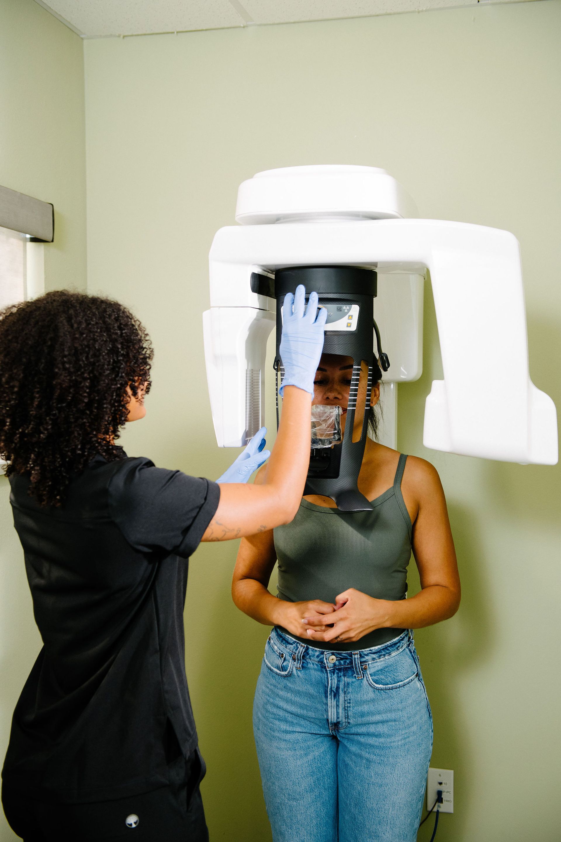
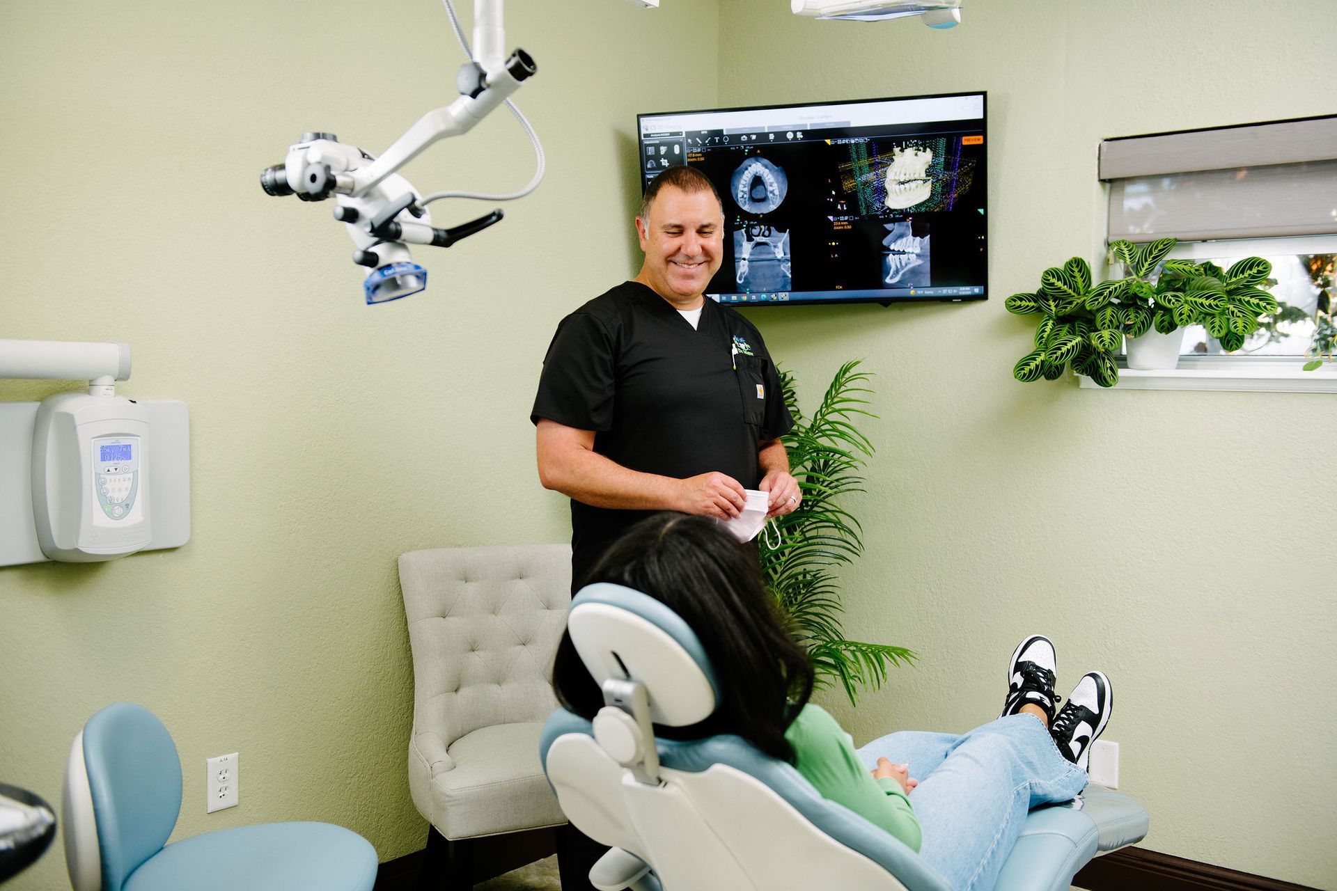
Dental radiographs are invaluable aids in diagnosing, treating, and maintaining dental health. Exposure time for dental radiographs is extremely minimal. Bayside Endodontics utilizes Digital Imaging Technologies within the office. With digital imaging, exposure time is about 90 percent less when compared to traditional radiographs. Digital imaging can also help us retrieve valuable diagnostic information. We may be able to see cavities better.


Digital imaging allows us to store patient images, and enables us to quickly and easily transfer them to specialists or insurance companies.

  • Digital X-Rays

    Introduced in 1987, nearly 90 years after traditional x-rays came to fruition, digital radiography combined the power of computer technology with electric sensors and tiny bursts of radiation. Rather than printing the results on film, images form almost as soon as the sensors are placed in our mouths, projecting on a computer screen. Digital x-ray technology does demand additional training for orthodontists, though the majority of practitioners are adamant that the advantages are worth the commitment. Today, a lot of orthodontic offices only offer patients digital x-rays because, in multiple ways, it is the superior option to traditional radiography.


    • Less Expensive | Digital x-rays will generally cost you less than the traditional alternative because the cost of film to develop images for the latter adds up. In contrast, digital x-ray imaging projects right onto our computer.
    • Better Storage | Since these digital x-ray images are transferred to a computer system, it allows for easier storage of your oral health records. Your data can be transferred from one practice to another without any medical data being lost in the exchange.
    • Finer Images | Digital x-ray images produce a better resolution than their traditional counterpart. Also, old-fashioned x-rays can only project images in 25 various shades, whereas a digital image can reveal up to 256 shades of grey. Digital radiography also has the advantage of accessing more angles within our mouths, providing a streamlined view of a patient's entire oral structure. With the assistance of computer programs, orthodontists can even enhance the digital images further, for a focused view.
  • CBCT Machine

    Cone beam computed tomography (CBCT) is a special type of x-ray machine that is implemented in scenarios where normal facial x-rays are insufficient. This variation of the CT scanner employs a special type of technology to generate 3D images of oral structures, soft tissues, nerve paths, and bone in the craniofacial area, in one scan. These images allow for more specific treatment planning. The CBCT machine has an x-ray beam, in the shape of a cone, which moves around you to create a large number of high-quality images, or views. It was developed as a means to produce similar images to what a CT provides, though with a significantly smaller and less costly machine that could be situated in an outpatient office. Providing detailed images of the bone, the CBCT machine evaluates diseases of the jaw, dentition, bony structures of the face, sinuses, and nasal cavity. One shortcoming is that it does not provide the comprehensive diagnostic information available with conventional CT, especially in the analyzing of soft tissue structures such as muscles, glands, nerves, and lymph nodes. The CBCT machine can also be used for reconstructive surgery, cephalometric analysis, locating the origin of pathology, surgical planning for impacted teeth, diagnosing temporomandibular joint disorder (TMD), and for the accurate placement of dental implants.

  • Surgical Microscopes

    The introduction of the surgical microscope has revolutionized the field of Endodontic Microsurgery. We have invested in the very best quality surgical microscopes, by Carl Zeiss, that provide unparalleled magnification and illumination for our surgical procedures.


    Our success depends on us being able to see the minutest of details – you cannot treat what you cannot see. The use of an endodontic microscope enhances the detection of root canals, reducing the likelihood of missed anatomy and contributing to more predictable and successful treatment outcomes.